
Management of community-associated bacterial skin and soft tissue infections in children and prevention of recurrence

Posted: May 26, 2026
Principal author(s)
Justin Penner MD, Sergio Fanella MD; Canadian Paediatric Society, Infectious Diseases and Immunization Committee
Abstract
Skin and soft tissue infections (SSTIs) are among the most common infectious diseases encountered in children. Severity of SSTIs range from simple infections that resolve spontaneously to severe necrotizing syndromes requiring intravenous antibiotics, surgical intervention, and intensive care. Inadequate skin barrier resulting from common paediatric conditions such as eczema and skin abrasions predisposes to infection. When antibiotics are needed, their choice and duration should be guided by the most probable organisms involved, their likely susceptibility profiles, and geographical epidemiologic differences. Inappropriate treatment with overly broad-spectrum antibiotics and/or for prolonged durations is detrimental to the microbiomes of patients, increases antimicrobial-related side effects, and perpetuates colonization of resistant organisms, precipitating antimicrobial resistance. This statement provides health care providers with a toolkit for optimal management of bacterial SSTIs, including recurrent episodes. It does not comprehensively examine SSTI treatment principles in immunocompromised or burn patients due to the management complexities of both settings.
Keywords: Abscess; Cellulitis; Decolonization; MRSA; Skin infection
Background
The skin protects its host from infection by acting as a physical barrier. Damage to this barrier from a variety of processes, including atopic disease, skin irritants, xerosis, micro abrasions, ectoparasites (e.g., scabies, lice), trauma, surgery, viral skin infections (e.g., herpes-zoster/herpes simplex [HZV/HSV]), or hair/nail infections, increases vulnerability to infection.
Skin and soft tissue infections (SSTIs) are some of the most common infectious diseases encountered in paediatrics. A Canadian multicentre study of 12 remote/isolated communities estimated the prevalence of SSTIs in some sites at 36.8%, with 60.2% of the total population studied having received at least one antibiotic prescription in the preceding 12 months[1]. Such high antimicrobial use may be linked to higher rates of antibiotic-resistant infections in these communities. Both the frequency of SSTIs in the paediatric population and the potential for high antimicrobial use resulting in infections with resistant organisms make antimicrobial stewardship a public health issue of paramount concern. This statement provides health care providers with a toolkit for optimizing the management of bacterial SSTIs, including recurrent episodes. It replaces previous guidance on methicillin-resistant Staphylococcus aureus (MRSA) skin abscesses from the Canadian Paediatric Society[2].
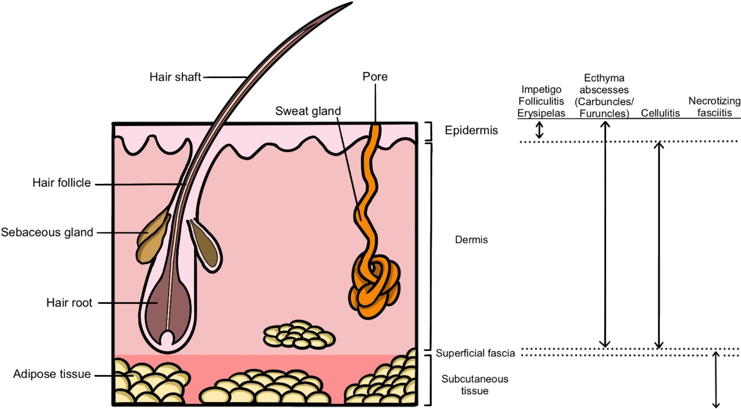
SSTIs have been categorized in various ways, without universal consensus. A list of common terms can be found in Table 1. Bacterial infections of the epidermis are typically less severe than deeper infections, but they can facilitate bacterial entry into deeper tissues. Abscesses can manifest in any of the tissue layers and are characterized by discrete collections of pus (Figure 1).
Table 1. Skin and soft tissue infections: Terms and definitions*
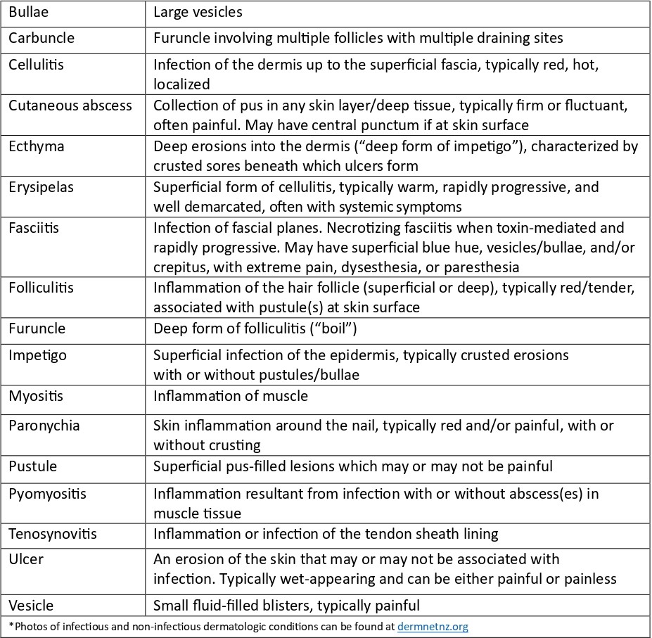
Figure 1. Skin and soft tissue infection anatomy
Delays in initiating treatment and inconsistencies in management
practices can cause disease progression, pain, deep-tissue abscesses,
musculoskeletal infections, and hypo- and hyperpigmentation.
Inappropriate antibiotic initiation, duration, and drug or route
selection for simple infections are detrimental to the individual’s
microbiome, increase antimicrobial-related side effects (e.g., Clostridioides difficile), and have societal impacts through increased antimicrobial resistance.
Microbiology
The most common pathogens associated with community-acquired bacterial SSTIs include methicillin-susceptible or -resistant Staphylococcus aureus (MSSA or MRSA) and Streptococcus pyogenes (Group A Streptococcus [GAS]). Both pathogens can cause a range of SSTIs, from mild infections (e.g., folliculitis, impetigo) to severe necrotizing infections complicated by toxic shock or sepsis. Recent reports have suggested an increase in invasive GAS infections[3]. Non-pyogenic cellulitis has been reported more with GAS compared with S. aureus, although microbiological confirmation is rare in this context and Canadian data is lacking in paediatric populations[4].
Although variable by province/territory, an increase in paediatric MRSA has been recognized in Canada[5][6]. This rise disproportionately impacts certain minoritized groups, such as Indigenous communities in remote/isolated areas, where MRSA proportions can exceed 50%[7]. Such prevalence indicates that certain social determinants of health, such as crowded housing, are risk factors for SSTIs[8]. Colonization rates of up to 67% have been reported in households where one member has had a recent MRSA infection[9]. Certain behaviours, such as nose-picking, have been associated with increasing rates of S. aureus carriage[10][11]. Some studies have shown that approximately one-third of individuals colonized with MRSA will develop an SSTI[12][13], and up to 70% with an MRSA SSTI may experience recurrence(s) over the following 6 to 12 months[14]. SSTI risk in children also increases with MSSA colonization[15].
Other bacterial pathogens and non-infectious mimics should be considered based on exposure history and examination. Waterborne pathogens include Aeromonas spp (in fresh water), Vibrio spp (in brackish or salt water), and Pseudomonas aeruginosa (causing hot tub folliculitis, or ecthyma). Anaerobic bacteria can cause SSTIs following a deep puncture wound or animal or human bite, and may feature in cases of longstanding ulcerative lesions, immunocompromised hosts, and prolonged or recent antimicrobial use. Consider non-tuberculous mycobacteria, candida, moulds, and cutaneous dermatophytes when the epidemiology or morphology of lesions suggests such etiologies. VZV infections predispose to SSTIs, including invasive infections (e.g., necrotizing fasciitis)[16]. Eczema can also be superinfected with HSV (eczema herpeticum) or enteroviruses (eczema coxsackium). Non-infectious mimics of SSTIs include, but not limited to: pyoderma gangrenosum, pyogenic granuloma, psoriasis, non-infected atopic dermatitis, hidradenitis suppurativa, venous stasis or thrombosis, vasculitis, urticaria, contact dermatitis, malignancy, and other non-infectious dermatologic or rheumatologic conditions.
History and physical examination
The epidemiological triad comprises the host, pathogen, and environment. A focused history should consider interactions among the three, including preceding skin trauma, bites (animal or human), water exposures, travel, sick contacts, social, occupational, and recreational activities, and hospitalization(s) or frequent health care admissions or attendance. Chronicity (i.e., acute, subacute, or chronic) and associated symptoms (i.e., fever, pain) should form part of an SSTI history. Past medical history should include personal and family experience of previous SSTIs, underlying medical conditions, known history or risk factors for MRSA colonization, and vaccination status. While most children with recurrent SSTIs are immunocompetent, a careful history should ensure there are no “red flags” for inborn errors of immunity, such as recurrent, invasive, multi-site, or unusual infections, early age of onset, deep skin or lymph node abscesses, failure to thrive, and severe or intractable eczema. In cases of recurrent perianal abscesses, inflammatory bowel disease should be considered.
Severity assessments are largely subjective, though scoring systems have been validated to help define mild, moderate, and severe disease. One scoring system for cellulitis is the Melbourne ASSET score. Higher scores suggest increasing severity, with a cut-off of 4 limiting unnecessary intravenous (IV) antibiotics[17]. In a case-based survey of paediatricians, the most common findings associated with severity and treatment with IV versus oral antimicrobials included tracking lymphangitis, functional impairment of the affected area, fever, oral antibiotics within the previous 24 hours, size, site, and degree of swelling and tenderness[18]. Further findings suggestive of severe invasive disease include pain out of proportion to clinical findings, rapidly expanding erythema, necrosis, and crepitus.
Diagnostics
Microbiological samples should be sought for open wounds or lesions with visible pus or fluid. Pus collected by needle aspirate, surgical, or manual drainage of abscesses should be cultured and prioritized over swabs based on their higher microbiological yield. Superficial swabs of intact and non-intact skin or ulcers are of limited use due to isolation of colonizing microbiota. If debridement is indicated, tissue samples for culture are of highest yield. Other samples (e.g., skin biopsy) may be considered in settings of chronic infection, failed therapy, an immunocompromised child, or unusual exposure(s). In such cases, an infectious disease (ID) or dermatology consult (or both) should be sought to guide specimen requests. Specialized viral swabs from deroofed vesicles placed in dedicated media are warranted for HSV, VZV, and enterovirus polymerase chain reaction (PCR) testing. Blood cultures are rarely indicated with clinically mild to moderate SSTIs but should be collected with systemic illness, shock, and severe or deep-seated infections. Dermatophyte and ectoparasite skin scrapings should be collected when clinically indicated, following local laboratory protocols.
Management
(Figure 2)
Medical management
Clinicians should first consider whether antimicrobials are warranted. Minor infections can resolve spontaneously with wound care alone. Best practices for wound care management can be found at woundscanada.ca. In addition to wound care, a short-course prescription (3 to 5 days) of topical antimicrobials may be required. Evidence of benefit for over-the-counter topical antimicrobials (e.g., polymyxin B, bacitracin, neomycin) is unclear[19]. Empiric antimicrobial choices should be individualized based on history (e.g., exposures), examination findings, and previous microbiology results when available. More recent microbiological results should be prioritized. Local paediatric-specific antibiograms are encouraged to help guide empiric antimicrobial choice.
Empiric antibiotics should target the most common pathogens causing SSTIs (Table 2[20]-[22]) and most infections are amenable to oral treatment. In the majority of cases, including moderate infections, oral cephalexin is appropriate, provided that adequate dosing is administered[23]. Combination therapy with cephalexin and trimethoprim/sulfamethoxazole or broad-spectrum outpatient IV antibiotics (e.g., ceftriaxone) are discouraged. Empiric therapy targeting MRSA may be warranted in some cases, such as when children are colonized or living in MRSA-prevalent areas (i.e., where >10% to 15% S. aureus isolates are MRSA)[24]. Oral trimethoprim/sulfamethoxazole is preferred due to high susceptibility rates for MRSA in Canada. Clindamycin resistance is higher, although this antibiotic may still be considered for children with prior susceptible isolates or when antibiograms demonstrate high susceptibility. When a patient is known to be carrying MRSA (e.g., MRSA-positive nares) or later found to be after initiating antimicrobials but has improved with non-MRSA treatment, switching antimicrobials is not necessary. For children colonized with MRSA or living in highly MRSA-endemic areas and presenting with a non-purulent SSTI (e.g., cellulitis) without systemic features, consider initiating a non-MRSA antimicrobial (e.g., cephalexin) with close follow-up[25][26].
For hospitalized children, antimicrobial choice is guided by the history and severity of disease. For non-toxic children who have not been exposed to atypical pathogens, IV cefazolin or, when MRSA is suspected, vancomycin, is appropriate. For children who are systemically unwell or toxic (e.g., concern for sepsis, fasciitis/myositis, shock), many experts recommend initiating broad-spectrum coverage (ceftriaxone or piperacillin-tazobactam, plus vancomycin) empirically, then discontinuing vancomycin when MRSA is excluded. The addition of an anti-toxin antibiotic (e.g., clindamycin) should be strongly considered in such cases[27]. ID consultation should be sought and antibiotics targeted to microbiology results. When SSTIs are complicated by a herpesvirus (e.g., HSV/VZV), IV acyclovir or oral acyclovir/valacyclovir may be warranted.
Table 2. Antimicrobials for treating systemic skin and soft tissue infections (SSTIs)

* Dose adjustments may be required based on hepatic/renal function or for infants. Consult with pharmacy as required.
† Low risk: <10%–15% S. aureus isolates are MRSA; High risk: >10%–15% S. aureus isolates are MRSA GAS Group A streptococcus; IV Intravenous; MRSA Methicillin-resistant Staphylococcus aureus; MSSA Methicillin-susceptible Staphylococcus aureus; TMP/SMX Trimethoprim/sulfamethoxazole
Surgical management
Source control of abscesses through surgical or manual drainage is vital. Warm compresses can facilitate drainage. When drainage of an uncomplicated small collection is achieved in a healthy host, without significant surrounding cellulitis, antimicrobials are rarely warranted[28]-[30]. If antimicrobials were started previously, they can typically be discontinued when drainage is achieved. Post-drainage wound care is important for healing and consultation with experts in wound care may be considered.
In cases of necrotizing infection, urgent surgical consultation for debridement is essential. Deep samples of pus and tissue should be sent for aerobic and anaerobic cultures. Debridement of chronic ulcers and biopsies of unusual or chronic lesions and those failing therapy ensures that tissue can be sent for bacterial, mycobacterial, and fungal cultures and for histopathology to aid diagnosis and targeted therapy.
Figure 2. Treatment algorithm for skin and soft tissue infections (SSTIs)
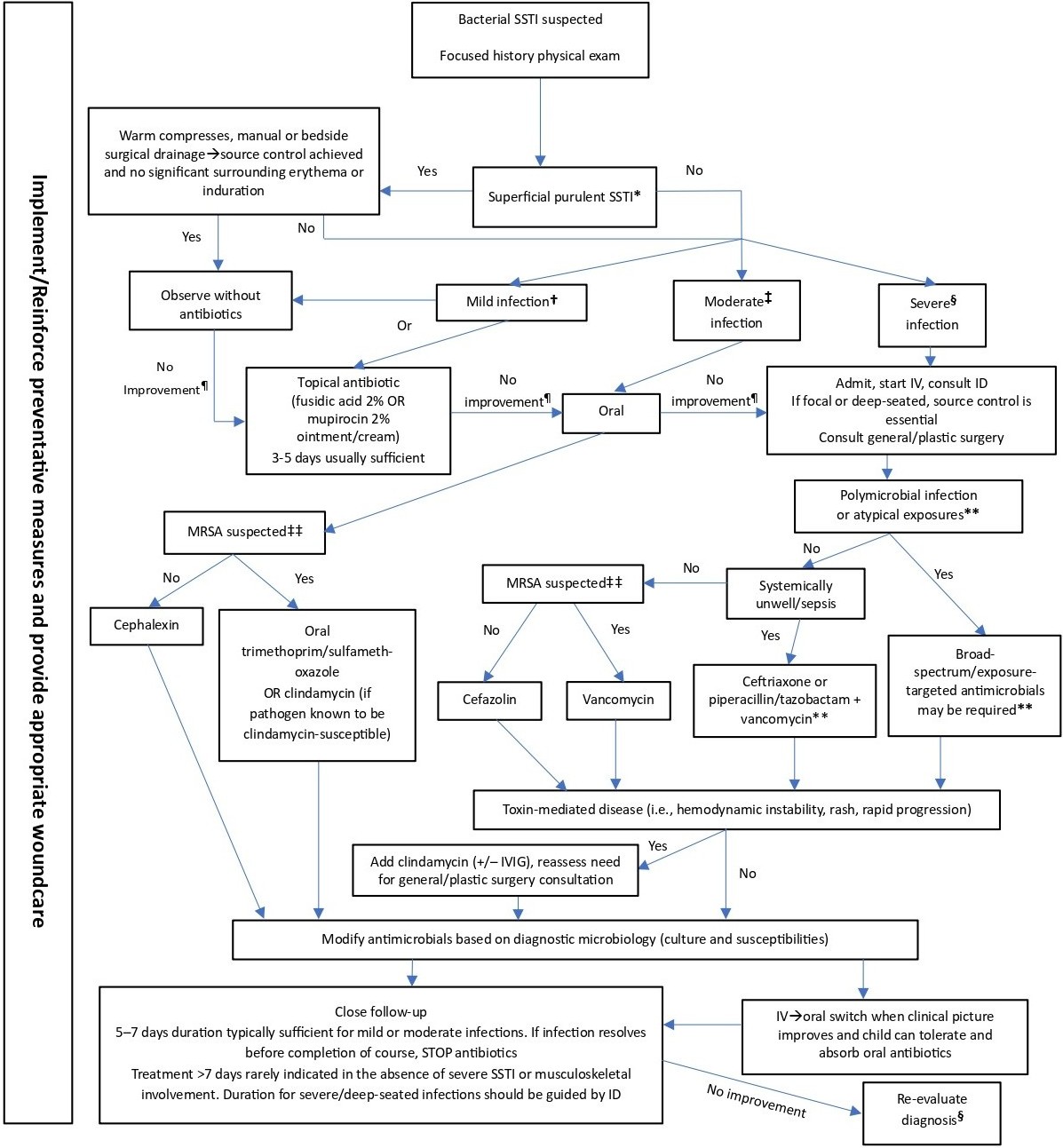
* Abscess, carbuncle, furuncle
† Mild infection (small area): Systemically well,
localized SSTI, no or minimal erythema or induration, no deep-seated
infection. Outpatient treatment. May resolve with appropriate skin care
alone
‡ Moderate infection (e.g., limited cellulitis): Systemically well, not
rapidly progressive, no deep-seated infection. Outpatient treatment
§ Severe infection (e.g., widespread
cellulitis/deep-seated infection): Extensive infection, clinically
unstable, rapidly progressive, necrosis, pain out of proportion,
crepitus. Inpatient treatment. If systemically unwell, start empiric
antibiotics urgently
¶ Re-evaluate diagnosis (repeat history/examination), need for source control
** Consult ID, refer to local antimicrobial guidelines
‡‡ MRSA risk factors: MRSA colonized, previous or recurrent MRSA SSTIs, local prevalence >10% to 15%
ID Infectious diseases; IV Intravenous; IVIG Intravenous immunoglobulin; MRSA Methicillin-resistant Staphylococcus aureus
Preventing recurrence
Preserving an intact skin barrier, including wound management and control of primary dermatologic disorders, is important to prevent SSTIs, including recurrence. Optimize eczema management, including breaking the pruritus→scratch→skin breakdown cycle. Dermatology consultation should be considered for children with difficult-to-control skin conditions that threaten recurrence or serious infection.
In healthy patients, decolonization has achieved mixed results in preventing SSTI recurrence, and treatment regimens and practices vary with no clear consensus[31]-[36]. Decolonization practices can also be costly and dry the skin if concurrent moisturizers are not used. Recurrence of colonization is common, even with diligent decolonization practices[37]-[39]. Some experts reserve decolonization for recurrence of moderate to severe SSTIs, household outbreaks, or to prepare for procedures with high infection risk. Challenging cases often benefit from ID consultation, and the advantages and disadvantages of treatments and their variable success rates should be discussed with families. When recommended, a 5-day trial of 2% mupirocin ointment twice daily to nares, combined with daily cleaning with 4% chlorhexidine soap or 2% chlorhexidine wipes (for 5 to 7 days) is suggested[40]. Dilute bleach baths (DBBs) are an acceptable alternative and can be used as part of both decolonization regimens (daily for 5 to 7 days) or maintenance suppression (or both)[41]. Concurrent family/household member decolonization may also be considered to increase chances of decolonization success, although evidence for this practice is inconsistent[42]-[44]. Use of oral antimicrobial agents for decolonization is discouraged unless directed by ID[31].
DBBs are prepared with 1/4 to 1/2 cup bleach added to a bathtub of water. Children should be encouraged to sit in the bath for 5 to 10 minutes. Swimming in a chlorinated pool has similar effect. Maintenance DBBs for children with a recurrent SSTI may be continued once or twice per week to suppress bacterial microbiota. Make sure that skin moisturizer is being applied after bathing. Concurrent environmental decolonization measures in the household include cleaning surfaces with bleach-containing solution, focusing on “high-touch” surfaces, washing towels, clothing, and bedding in hot water, and replacing or disinfecting personal hygiene items[45]-[47]. Sharing such items and sharing athletic equipment are discouraged. To help maximize success, clinicians are encouraged to develop written handouts describing these regimens, including images, detailed instructions, with translation into local languages.
Additional measures to decrease recurrence include diligent wound care, covering open wounds, encouraging universal uptake of varicella vaccination, and regular cleaning of communal, sporting, and exercise equipment (ideally after each use). Scabies and lice predispose to SSTIs and should be treated per local guidelines[48][49]. Chronic antibiotic suppression is rarely indicated but may be considered in consultation with ID for certain inborn errors of innate immunity, chronic or recurrent dermatologic diseases, or conditions of aberrant lymphatic drainage.
Recommendations
- Managing skin and soft tissue infections (SSTIs) should be based on a focused history and careful assessment of the severity of infection.
- Empiric antimicrobial prescribing for SSTIs should be based on locally sourced and paediatric-specific antibiograms. Prioritize the most narrow-spectrum antimicrobial.
- Manage minor SSTIs with drainage and/or skin care alone. In select cases, the addition of topical antimicrobial treatment may be required.
- Adequate drainage of abscesses negates the need to initiate antimicrobials in most outpatient cases. If antimicrobials are commenced, they can be discontinued when source control is achieved, provided there is no significant surrounding cellulitis.
- When oral antimicrobials are warranted, cephalexin monotherapy is the antimicrobial of choice. When methicillin-resistant Staphylococcus aureus is suspected, trimethoprim/sulfamethoxazole monotherapy is the antimicrobial of choice for outpatient management. It also has activity against Group A streptococcal infections.
- Short courses of antimicrobials (5 to 7 days) are often sufficient. Longer treatment durations should be reserved for more severe or complicated infections. Even severe SSTIs rarely require >7 days of antimicrobials. Failure to respond should prompt re-evaluation of the diagnosis and assessment for source control.
- Reserve intravenous antimicrobials for severe, deep-seated, or necrotizing infections. Narrow-spectrum, short-courses of antimicrobials should be prioritized based on microbiological testing, with a focus on source control.
- Preventing recurrent SSTIs should focus on maintenance of intact skin barrier, treatment of underlying skin conditions (e.g., eczema), and good wound and abrasion care.
- In select individuals and households, decolonization protocols can be considered.
Acknowledgement
This position statement was reviewed by the Community Paediatrics and First Nations, Inuit and Métis Health Committees of the Canadian Paediatric Society (CPS). It was also reviewed by the CPS Community Paediatrics, Hospital Paediatrics, and Paediatric Emergency Medicine Section Executives, and by members of the Association of Medical Microbiology and Infectious Disease Canada (AMMI).
CANADIAN PAEDIATRIC SOCIETY INFECTIOUS DISEASES AND IMMUNIZATION COMMITTEE (2024-2025)
Members: Michelle Barton MD (Chair), Laura Sauvé MD
(Past Chair), Eugene Ng MD (Board Representative), Ari Bitnun MD,
Jeannette Comeau MD MSc, Sergio Fanella MD, Justin Penner MD, MSc
Liaisons: Dorothy Moore MD (National Advisory Committee
on Immunization), Ari Bitnun MD (Canadian Paediatric and Perinatal
HIV/AIDS Research Group), Isabelle Viel-Thériault MD (Committee to
Advise on Tropical Medicine and Travel), Marina Salvadori MD (Public
Health Agency of Canada), Sean O’Leary (American Academy of Pediatrics,
Committee on Infectious Diseases), Rupeena Purewal MD (Immunization
Monitoring Program, ACTive), Cora Constantinescu MD (Association of
Medical Microbiology and Infectious Disease Canada, Pediatric Committee)
Principal authors: Justin Penner MD, Sergio Fanella MD
Funding
There is no funding to declare.
Potential Conflict of Interest
Dr. Fanella reported receiving funding as a local site PI for MCT for
COVID19 vaccines in children (ModernaTx). No other disclosures were
reported.
References
- Jeong D, Nguyen HNT, Tyndall M, Schreiber YS. Antibiotic use among twelve Canadian First Nations communities: A retrospective chart review of skin and soft tissue infections. BMC Infect Dis 2020;20(1):118. doi: 10.1186/s12879-020-4842-1
- Robinson JL, Salvadori MI; Canadian Paediatric Society, Infectious Diseases and Immunization Committee. Management of community-associated methicillin-resistant Staphylococcus aureus skin abscesses in children. Paediatr Child Health 2011;16(2):115-6. doi: 10.1093/pch/16.2.115
- Centers for Disease Control and Prevention. Group A Strep Surveillance and Trends. July 8, 2024 (Accessed October 22, 2025).
- Stevens DL, Bryant AE. Impetigo, Erysipelas and Cellulitis. February 10, 2016. In: Ferretti JJ, Stevens DL, Fischetti VA, eds. Streptococcus pyogenes: Basic Biology to Clinical Manifestations. Oklahoma City, OK: University of Oklahoma Health Sciences Center; 2016.
- Nichol KA, Adam HJ, Golding GR, et al; Canadian Antimicrobial Resistance Alliance (CARA) and CANWARD. Characterization of MRSA in Canada from 2007 to 2016. J Antimicrob Chemother 2019;74(Suppl 4):iv55-iv63. doi: 10.1093/jac/dkz288
- Public Health Agency of Canada. Canadian Antimicrobial Resistance Surveillance System (CARSS) Report 2022. January 2023 (Accessed October 22, 2025).
- Fanella S, Embree J. Pediatric Staphylococcus aureus infections: Impact of methicillin resistance at a Canadian center. South Med J 2015;108(5):254-7. doi: 10.14423/SMJ.0000000000000274
- Butler-Jones D, Wong T. Infectious disease, social determinants and the need for intersectoral action. Can Commun Dis Rep 2016;42(Suppl 1):S118-S120.
- Fritz SA, Hogan PG, Hayek G, et al. Staphylococcus aureus colonization in children with community-associated Staphylococcus aureus skin infections and their household contacts. Arch Pediatr Adolesc Med 2012;166(6):551-7. doi: 10.1001/archpediatrics.2011.900
- Loewen K, Schreiber Y, Kirlew M, Bocking N, Kelly L. Community-associated methicillin-resistant Staphylococcus aureus infection: Literature review and clinical update. Can Fam Physician 2017;63(7):512-20. Erratum in: Can Fam Physician 2017;63(8):596.
- Wertheim HFL, van Kleef M, Vos MC, Ott A, Verbrugh HA, Fokkens W. Nose picking and nasal carriage of Staphylococcus aureus. Infect Control Hosp Epidemiol 2006;27(8):863-7. doi: 10.1086/506401
- Ellis MW, Hospenthal DR, Dooley DP, Gray PJ, Murray CK. Natural history of community-acquired methicillin-resistant Staphylococcus aureus colonization and infection in soldiers. Clin Infect Dis 2004;39(7):971-9. doi: 10.1086/423965
- Yang ES, Tan J, Eells S, Rieg G, Tagudar G, Miller LG. Body site colonization in patients with community-associated methicillin-resistant Staphylococcus aureus and other types of S. aureus skin infections. Clin Microbiol Infect 2010;16(5):425-31. doi: 10.1111/j.1469-0691.2009.02836.x
- Creech CB, Al-Zubeidi DN, Fritz SA. Prevention of recurrent staphylococcal skin infections. Infect Dis Clin North Am 2015;29(3):429-64. doi: 10.1016/j.idc.2015.05.007
- Faden H, Lesse AJ, Trask J, et al. Importance of colonization site in the current epidemic of staphylococcal skin abscesses. Pediatrics 2010;125(3):e618-24. doi: 10.1542/peds.2009-1523
- Davies HD, McGeer A, Schwartz B, et al. Invasive group A streptococcal infections in Ontario, Canada. Ontario Group A Streptococcal Study Group. N Engl J Med 1996;335(8):547-54. doi: 10.1056/NEJM199608223350803
- Ibrahim LF, Hopper SM, Donath S, Salvin B, Babl FE, Bryant PA. Development and validation of a cellulitis risk score: The Melbourne ASSET Score. Pediatrics 2019;143(2):e20181420. doi: 10.1542/peds.2018-1420
- Ibrahim LF, Babl FE, Hopper SM, Bryant PA. Cellulitis: Oral versus intravenous and home versus hospital—What makes clinicians decide? Arch Dis Child 2020;105(4):413-15. doi: 10.1136/archdischild-2019-316824
- Banerjee S, Argáez C. Topical antibiotics for infection prevention: A review of the clinical effectiveness and guidelines. Ottawa, Ont.: Canadian Agency for Drugs and Technologies in Health; March 30, 2017.
- Kimberlin DW, Banerjee R, Barnett ED, Lynfield R, Sawyer MH. Tetracyclines. In: Red Book: 2024–2027 Report of the Committee on Infectious Diseases (33rd edn.). Itaska, IL: American Academy of Pediatrics; May 2024.
- Ravindra D, Huang G, Hallett K, Burgner DP, Gwee A, Silva MJ. Antibiotic exposure and dental health: A systematic review. Pediatrics 2023;152(1):2023061350. doi: 10.1542/peds.2023-061350
- McCreary EK, Johnson MD, Jones TM, et al. Antibiotic myths for the infectious diseases clinician. Clin Infect Dis 2023;77(8):1120-5. doi: 10.1093/cid/ciad357. Erratum in: Clin Infect Dis 2024;78(6):1780.
- Trottier ED, Farley St-Amand B, Vincent M, et al. Outpatient management of moderate cellulitis in children using high-dose oral cephalexin. Paediatr Child Health 2022;27(4):213-9. doi: 10.1093/pch/pxac031
- Kaplan SL. Treatment of community-associated methicillin-resistant Staphylococcus aureus infections. Pediatr Infect Dis J 2005;24(5):457-8. doi: 10.1097/01.inf.0000164162.00163.9d
- Eells SJ, Chira S, David CG, Craft N, Miller LG. Non-suppurative cellulitis: Risk factors and its association with Staphylococcus aureus colonization in an area of endemic community-associated methicillin-resistant S. aureus infections. Epidemiol Infect 2011;139(4):606-12. doi: 10.1017/S0950268810001408
- Rajan S. Skin and soft-tissue infections: Classifying and treating a spectrum. Cleve Clin J Med 2012;79(1):57-66. doi: 10.3949/ccjm.79a.11044
- Farrell CA. Diagnosis and management of severe sepsis in the paediatric patient. Paediatr Child Health 2020;25(7):475-6. doi: 10.1093/pch/pxz178
- Singer AJ, Thode HC. Systemic antibiotics after incision and drainage of simple abscesses: A meta-analysis. Emerg Med J 2014;31(7):576–8. doi: 10.1136/emermed-2013-202571
- Daum RS, Miller LG, Immergluck L, et al. A placebo-controlled trial of antibiotics for smaller skin abscesses. N Engl J Med 2017;376(26):2545–55. doi: 10.1056/NEJMoa1607033
- Wang W, Chen W, Liu Y, et al. Antibiotics for uncomplicated skin abscesses: Systematic review and network meta-analysis. BMJ Open 2018;8(2):e020991. doi: 10.1136/bmjopen-2017-020991
- Helbo T, Boel JB, Bartels MD, Ahlström MG, Holzknecht BJ, Eriksen HB. Carriage of methicillin-resistant Staphylococcus aureus in children <6 years old: A retrospective follow-up study of the natural course and effectiveness of decolonization treatment. J Antimicrob Chemother 2024;79(4):826-34. doi: 10.1093/jac/dkae036
- Piewngam P, Otto M. Staphylococcus aureus colonisation and strategies for decolonisation. Lancet Microbe 2024;5(6):e606-18. doi: 10.1016/S2666-5247(24)00040-5
- Klempner MS, Styrt B. Prevention of recurrent staphylococcal skin infections with low-dose oral clindamycin therapy. JAMA 1988;260(18):2682-5.
- Raz R, Miron D, Colodner R, Staler Z, Samara Z, Keness Y. A 1-year trial of nasal mupirocin in the prevention of recurrent staphylococcal nasal colonization and skin infection. Arch Intern Med 1996;156(10):1109-12.
- Coates T, Bax R, Coates A. Nasal decolonization of Staphylococcus aureus with mupirocin: Strengths, weaknesses and future prospects. J Antimicrob Chemother 2009;64(1):9-15. doi: 10.1093/jac/dkp159
- Creech CB, Beekmann SE, Chen Y, Polgreen PM. Variability among pediatric infectious diseases specialists in the treatment and prevention of methicillin-resistant Staphylococcus aureus skin and soft tissue infections. Pediatr Infect Dis J 2008;27(3):270-2. doi: 10.1097/INF.0b013e31815c9068
- Doebbeling BN, Reagan DR, Pfaller MA, Houston AK, Hollis RJ, Wenzel RP. Long-term efficacy of intranasal mupirocin ointment. A prospective cohort study of Staphylococcus aureus carriage. Arch Intern Med 1994;154(13):1505-8. doi: 10.1001/archinte.1994.00420130101013
- Fernandez C, Gaspar C, Vindel AT, Saez-Nieto JA, Cruzet F, Aguilar L. A double-blind, randomized, placebo-controlled clinical trial to evaluate the safety and efficacy of mupirocin calcium ointment for eliminating nasal carriage of Staphylococcus aureus among hospital personnel. J Antimicrob Chemother 1995;35(3):399-408. doi: 10.1093/jac/35.3.399
- Mody L, Kauffman CA, McNeil SA, Galecki AT, Bradley SF. Mupirocin-based decolonization of Staphylococcus aureus carriers in residents of 2 long-term care facilities: A randomized, double-blind, placebo-controlled trial. Clin Infect Dis 2003;37(11):1467-74. doi: 10.1086/379325
- Whitman TJ, Herlihy RK, Schlett CD, et al. Chlorhexidine-impregnated cloths to prevent skin and soft-tissue infection in Marine recruits: A cluster-randomized, double-blind, controlled effectiveness trial. Infect Control Hosp Epidemiol 2010;31(12):1207-15. doi: 10.1086/657136
- Fisher RG, Chain RL, Hair PS, Cunnion KM. Hypochlorite killing of community-associated methicillin-resistant Staphylococcus aureus. Pediatr Infect Dis J 2008;27(10):934-5. doi: 10.1097/INF.0b013e318175d871
- Fritz SA, Hogan PG, Hayek G, et al. Household versus individual approaches to eradication of community-associated Staphylococcus aureus in children: A randomized trial. Clin Infect Dis 2012;54(6):743-51. doi: 10.1093/cid/cir919
- Wiese-Posselt M, Heuck D, Draeger A, et al. Successful termination of a furunculosis outbreak due to lukS-lukF-positive, methicillin-susceptible Staphylococcus aureus in a German village by stringent decolonization, 2002-2005. Clin Infect Dis 2007;44(11):e88-95. doi: 10.1086/517503
- Hogan PG, Parrish KL, Mork RL, et al. HOME2 Study: Household versus personalized decolonization in households of children with methicillin-resistant Staphylococcus aureus skin and soft tissue infection—A randomized clinical trial. Clin Infect Dis 2021;73(11):e4568-77. doi: 10.1093/cid/ciaa752
- Hogan PG, Mork RL, Thompson RM, et al. Environmental methicillin-resistant Staphylococcus aureus contamination, persistent colonization, and subsequent skin and soft tissue infection. JAMA Pediatr 2020;174(6):552-62. doi: 10.1001/jamapediatrics.2020.0132
- Huang SS, Singh R, McKinnell JA, et al; Project CLEAR Trial. Decolonization to reduce postdischarge infection risk among MRSA carriers. N Engl J Med 2019;380(7):638-50. doi: 10.1056/NEJMoa1716771
- Mork RL, Hogan PG, Muenks CE, et al. Longitudinal, strain-specific Staphylococcus aureus introduction and transmission events in households of children with community-associated meticillin-resistant S aureus skin and soft tissue infection: A prospective cohort study. Lancet Infect Dis 2020;20(2):188-98. doi: 10.1016/S1473-3099(19)30570-5
- Banerji A; Canadian Paediatric Society, First Nations, Inuit and Métis Health Committee. Scabies. Paediatr Child Health 2015;20(7):395-402. doi: 10.1093/pch/20.7.395
- Cummings C, Finlay JC, MacDonald NE. Head lice infestations: A clinical update. Paediatr Child Health 2018;23(1):e18-e24. doi: 10.1093/pch/pxx165
Disclaimer: The recommendations in this position statement do not indicate an exclusive course of treatment or procedure to be followed. Variations, taking into account individual circumstances, may be appropriate. Internet addresses are current at time of publication.


